Skip to content
Paper language: English
Paper language: Spanish
Paper language: English and spanish
screencapture-facerevolution-org-wp-content-uploads-2021-03-articulo-placas-de-ateroma-2frrht-pdf-1616697189339
Detección de placas de ateroma mediante radiografías dentales
AUTHOR: Aranzazu Senosiain Oroquieta
English abstract
Stroke is the third leading cause of death in the United States and almost thehalf of deaths in Spain. One of the possible causes of stroke is the formation of atheroma plaques inthe carotid bifurcation. Eventually, these plaques calcify and can be detected by chance on panora-mic and lateral cephalometric radiographs. Material and Methods: The panoramic radiographs of 1,300patients of the Dental School of the University of Oviedo, Spain, were evaluated. For our study, onlypatients over the age of 40 were analized, resulting in a final population of 459 patients (194 men and265 women). In the cases in which a lesion was suspected, the dental history of the patients was stu-died in order to determine the presence of risk factors related with atherosclerosis: tobacco, alcohol,prior stroke or ischemic attacks, hypertension. Results: 13 individuals (2,83% of the final study popu-lation), (53,84% men and 46,15% women) had radiopacities in the panoramic radiographs which wereclassified as atheroma plaques. Those lesions were measured in a greyscale and the results were verysimilar to those obtained in the retromolar alveolar bone. 30,76% of the positive cases, showed bila-teral radiopacities, and 69,23% unilateral ones. Conclusion: dentists must pay special attention to thepossible detection of atheroma plaques on panoramic and lateral cephalometric radiographs, speciallyin those patiens with high risk of stroke.
Spanish abstract
El accidente cerebrovascular o «stroke» es la tercera causa de muerte en Esta-dos Unidos y representa casi la mitad de las muertes ocurridas cada año en España. Una de las posi-bles causas del stroke es la formación de placas de ateroma en la bifurcación carotídea. En ocasiones,estas placas se calcifican y pueden ser detectadas de manera casual en la consulta dental medianteuna radiografía panorámica o telerradiografía. Material y método: se han revisado las ortopantomo-grafías de 1300 pacientes pertenecientes a la Escuela de Estomatología de la Universidad de Oviedo.Para el estudio, sólo se tuvieron en cuenta los pacientes mayores de 40 años, resultando una pobla-ción definitiva de 459 pacientes (194 hombres y 265 mujeres). En caso de sospecha de lesión, se estu-dió la historia clínica de dichos pacientes para determinar la presencia de otros factores de riesgo rela-cionados con la aterosclerosis, y se hizo una valoración aproximada de la densidad ósea de las mismas.Resultados: 13 individuos (2,83% de la población de estudio), (53,84% hombres y 46,15% mujeres) pre-sentaron radiopacidades en las ortopantomografías y telerradiografías que fueron clasificadas comoplacas de ateroma. Dichas lesiones, medidas en una escala de grises, registraron unos valores muy simi-lares al obtenido a nivel del hueso alveolar retromolar. De los 13 casos positivos, 30,76% presentaronlas lesiones bilateralmente, y 69,23% unilateralmente. Conclusión: el odontólogo deberá prestar espe-cial atención a la posible detección de placas de ateroma mediante radiografías panorámicas y tele-rradiografías, especialmente en aquellos pacientes con mayor riesgo de stroke.

Download paper (PDF)

Access our most valuable content free of charge.

Related Scientific papers

1st FACE online symposium

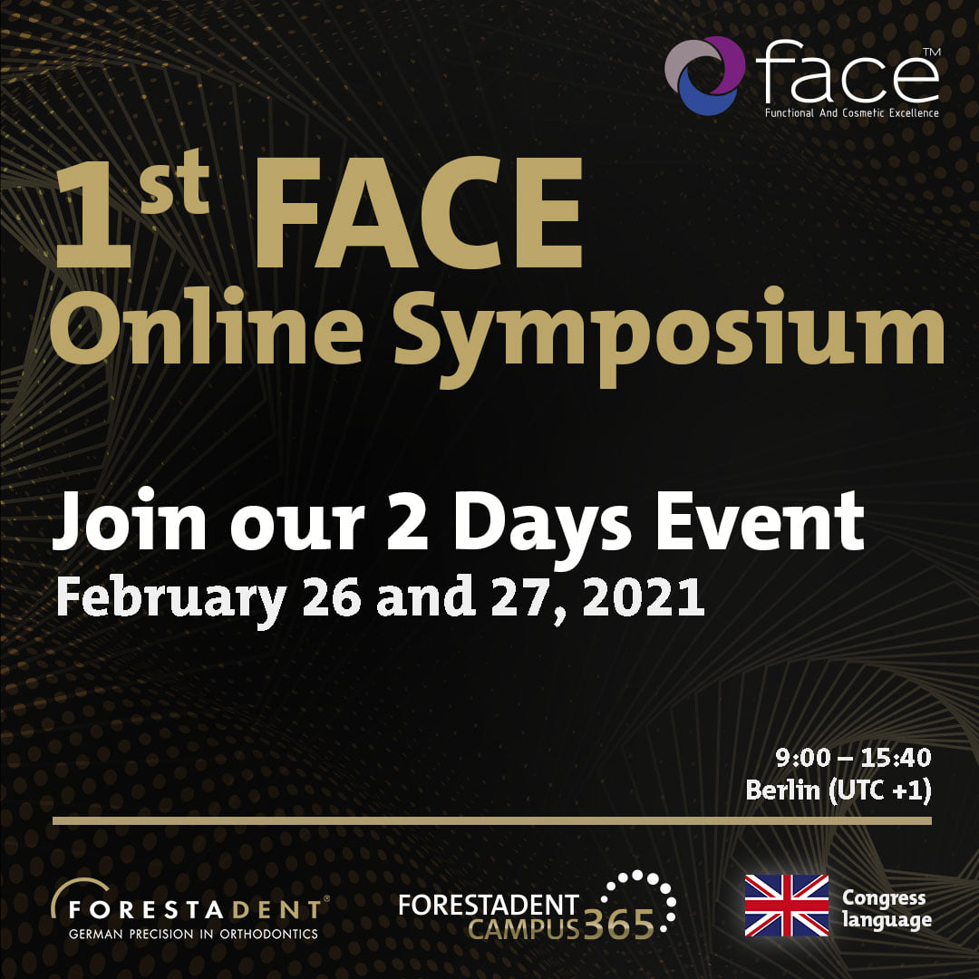
The world is changing and in FACE, following tradition, we wont be left behind.

As we all know, we can’t travel or meet, so once again, we will take advantage of technology to turn the situation around.

«Work hard, play hard«

Two days full of experiences, thanks to the participation of 20 different clinics.

We’ll see you on February 26 and 27